Quantitative Detection of Engineered Nanoparticles
in Tissues and Organs: An Investigation of Efficacy
and Linear Dynamic Ranges Using ICP-AES
Hans C. Fischer, Sebastien Fournier-Bidoz, K. Sandy Pang, Warren C. W. Chan
Nanobiotechnol (2007) 3:46-54 DOI 10.1007/s 12030-007-0006-2
Abstract
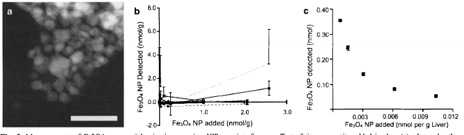
The absence of effective non-isotopic quantification methods to determine in vivo nanoparticle kinetics and distribution is a key obstacle to the development of various biomedical nanotechnologies. This paper presents a novel adaptation of the established technology of Inductively Coupled Plasma-Atomic Emission Spectroscopy (ICP-AES) to a simple technique intended to address this obstacle. Applicability to three varieties of nanoparticles, (CdSe/ZnS) quantum dots (QD), gold nanoparticles, and Fe304 nanopanicles) was investigated, and particle detection sensitivity was shown in moles of panicles per gram of tissue. Using gold nanoparticles, increased particle size corresponded with lower molar detection thresholds. Minimum linear detection ranges of 2.5 orders of magnitude for QDs and 1.5 orders of magnitude for all three sizes of gold were demonstrated. The detection of the Fe30 4 particles was hampered by the natural presence of Fe 2+ in tissues, showing that the technique is not suitable for measuring nanoparticles composed of endogenous elements. These detection levels and ranges demonstrate that this technique is useful for quantifying nanoparticles in excised organs, after in vivo dosing.