Transcribing In Vivo Blood Vessel Networks into In Vitro Perfusable Microfluidic Devices
Yih Yang Chen, Benjamin R Kingston, Warren CW Chan
Advanced Materials Technologies, Volume 5, Issue 6 | DOI:10.1002/admt.202000103
Abstract
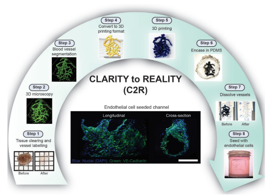
The 3D architecture of blood vessel networks dictates how nutrients, waste, and drugs are transported. These transport processes are difficult to study in vivo, leading researchers to develop methods to construct vessel networks in vitro. However, existing methods require expensive, customized equipment and cannot create large (>1 cm3) constructs. This makes them inaccessible to many researchers or educators. Here, a method that transcribes 3D images of blood vessel networks into physical microfluidic devices is developed. The method takes 3D images of blood vessel networks and uses fused‐filament 3D fabrication with standard polylactic acid (PLA) filament to print the imaged vessel network. The 3D printout is cast in polydimethylsiloxane (PDMS) and dissolved, producing vessel channels that are lined with endothelial cells. Devices imprinted with different vessel networks including small intestinal villi, pancreatic islets, and tumors from mice and humans are created. The method replicates the complex geometries of blood vessel networks in an in vitro device with commonly available equipment and materials. This increases the accessibility of this technology by allowing researchers or educators without access to expensive laser ablation microscope set‐ups or custom 3D printers to be able to create vasculature network devices.