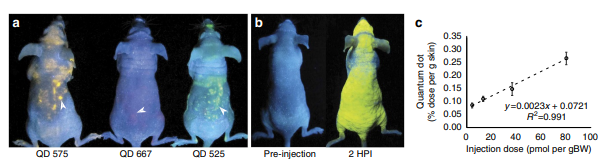
Nanoparticle exposure in animals can be visualized in the skin and analysed via skin biopsy
Edward A Sykes, Qin Dai, Kim M Tsoi, David M Hwang, Warren CW Chan
Nature Communications, volume 5, Article number: 3796 (2014) | DOI: 10.1038/ncomms4796
Abstract
The increasing use of nanomaterials raises concerns about the long-term effects of chronic nanoparticle exposure on human health. However, nanoparticle exposure is difficult to evaluate non-invasively using current measurement techniques. Here we show that the skin is an important site of nanoparticle accumulation following systemic administration. Mice injected with high doses of gold nanoparticles have visibly blue skin while quantum dot-treated animals fluoresce under ultraviolet excitation. More importantly, elemental analysis of excised skin correlates with the injected dose and nanoparticle accumulation in the liver and spleen. We propose that skin analysis may be a simple strategy to quantify systemic nanoparticle exposure and predict nanoparticle fate in vivo. Our results suggest that in the future, dermal accumulation may also be exploited to trigger the release of ultraviolet and visible light-sensitive therapeutics that are currently impractical in vivo due to limits in optical penetration of tissues at these wavelengths.